March 8, 2024

According to WHO, breast cancer is the most common cancer in the world and the 2nd most common cause of death. To assess the type of tumour and determine the most appropriate treatment, tissue is biopsied, stained and observed under a microscope (histological images). This is a lengthy process, requiring the know-how of an experienced pathologist. In partnership with Maria Vakalopoulou and Stergios Christodoulidis, professors at the Université de Paris-Saclay (CentraleSupélec), researchers at ÉTS and CNRS aim to automate the analysis of these tissues to speed up the work of pathologists.
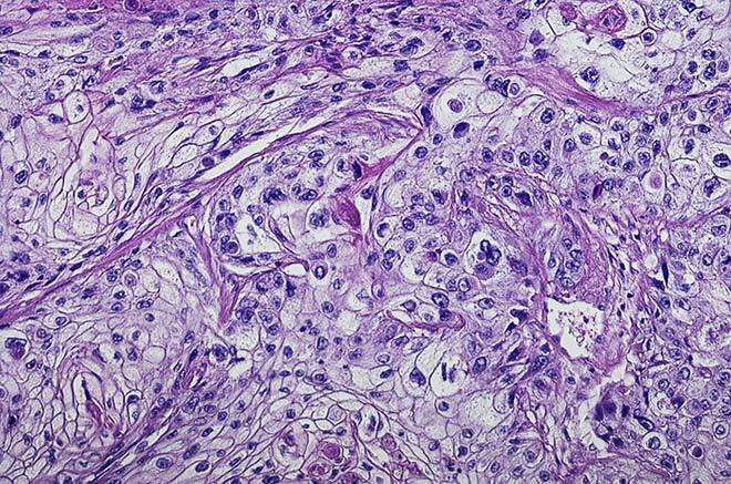
Figure 1 Histological image
Building on a Foundation Model
Deep learning models (artificial intelligence) are powerful tools for this kind of application, but they have their limitations. On the one hand, they require a lot of data (images) annotated by specialists for training, a time-consuming and costly task. On the other hand, histological images can vary significantly when taken under different conditions, and tissue staining protocols may differ from one pathologist to another. These changes lead to modifications in tissue images observed under the microscope and can skew their classification (domain change).
Our team has chosen to work with a more generalist foundation model, which will be trained to recognize general patterns in histological images from a large and varied database. This robust model can then be adapted to new tasks with little data, generally from one to five annotated examples for each new task.
Moreover, instead of just outlining the tumour, physician comments associated with each image will be included to add further details on the type and severity of tumours, as well as other characteristics observed in the images.
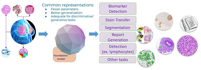
Figure 2 Approach
Better Predictors of Uncertainty
Before transferring our system to clinical practice, we need to be able to assess the degree of certainty in the algorithm predictions: one error can have a very serious impact on a woman’s life. At the same time, we intend to develop new learning strategies that will improve the degree of the certainty of the predictions. The aim of these strategies will be to detect when the algorithm encounters a problem and flag it so that a human can pay more attention to problematic images.
Lightening the Load on Specialists to Boost Productivity
This research project aims not to replace humans with algorithms but to make their work easier so that they can process more cases at a lower cost. Pathologists will be able to pay more attention to predictions classified as doubtful by the prediction evaluation tool.
The source code of our algorithm and the trained generalist models will be made available to the entire research community.