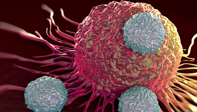
Electromagnetic Waves to Diagnose Cancer
Cancer screening tests take many forms. Some involve undergoing a magnetic resonance test while others involve tissue removal from various organs of the body. Other types of tests, such as the one for prostate cancer, measure the concentration of markers—proteins secreted by certain cancer cells—in the blood. Although this type of test is less invasive for the patient, it has two major drawbacks. First, the type of cancer suffered by the patient must be known in advance, so the test cannot be a blind one. Second, as some markers are not exclusive to a specific type of cancer, the use of markers is limiting.
Vahé Nerguizian, a professor in the Department of Electrical Engineering at the École de technologie supérieure (ÉTS), wants to facilitate diagnosis by developing a unit with MEMS (Micro Electro Mechanical Systems)/microfluidic sensors and an electromagnetic microwave device. Here is how the unit operates: after the red blood cells and the white blood cells have been removed (Figure 1), the blood passes through a microfluidic channel (Figure 2) where a high frequency electromagnetic wave, close to cellular wave frequency, is injected (Figure 3). For each cell passing through, the part of the signal penetrating it and the part that is reflected are measured and compared with other measurements from a big data bank (using a neural network system). Cancer cells are detected because of their dielectric properties, which are different from healthy cells.
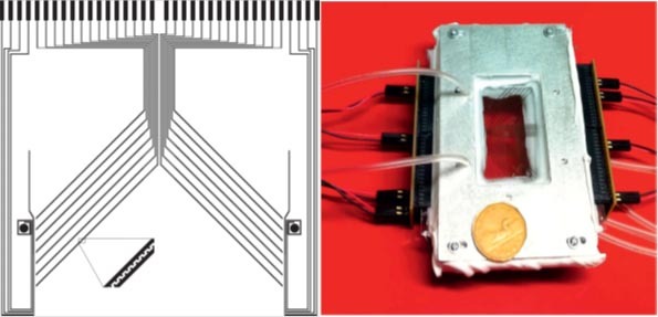
Figure 1 Separation system for red and white blood cells, and cancer cells via dielectrophoresis
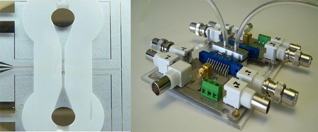
Figure 2 Microfluidic channel system with MEMS “antenna” sensors
This type of screening could be done during an annual check-up with the family physician. It would have the benefit of diagnosing cancers at an early stage and could identify people in need of further testing. It would serve as a “blind” detection for many forms of cancer and, as nothing is added to the blood, cancer cells could be recovered for further analysis, including an evaluation of the effectiveness of certain treatments such as chemotherapy. The uniqueness and innovation of this device lie in its ability to detect cancer without the use of detection markers.
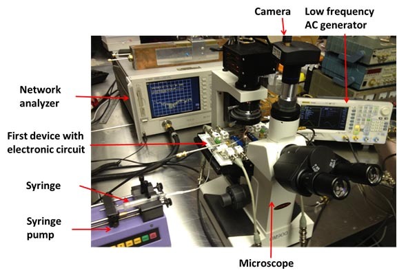
Figure 3 Microfluidic canal, microwave antenna, and electronic environment integrated system
Minimizing Prostate Biopsies
Another of Professor Nerguizian’s research projects is the development of a device to perform more accurate and less invasive prostate biopsies. The method currently used consists in taking samples from 10 to 16 sampling points on a grid determined by the doctor. The more samples taken, the longer the wounded prostate will take to heal. On the other hand, if the affected tissue area is small, the spot can be missed and existing cancer may not be detected.
The proposed device includes a miniature MEMS antenna connected to a very fine needle. This antenna will sweep the whole surface of the prostate. When it detects a change in the tissue’s dielectric properties, it pierces the area in order to take a sample. The advantages of this method are a systematic scan of the prostate and a smaller number of unnecessary sample extractions. However, the heterogeneity of prostate tissue presents a challenge that will have to be overcome.
A Liposome-Producing Factory
 Lastly, Professor Nerguizian is also working on a research project focusing on liposome production. Liposomes are artificial vesicles formed by lipid bilayers. Attached to them are markers and drugs that target cancer cells by reaching the desired location or cancer tissue. Specific repeatable (liposome sizes vary greatly) and industrial liposomes are difficult to produce: pressure and temperature must be precisely regulated. Professor Nerguizian wants to develop a technology that would produce stable liposomes in sizes that are well controlled in microfluidic channels.
Additional Information
Students interested in these research projects can contact Professor Vahé Nerguizian for more information.