A Predictive Model in Ultrasound Phantom Design
Purchased from Istock.com. Copyright.
Phantoms are devices that reproduce real life characteristics (mechanical, acoustic, etc.) in a controlled manner to validate medical imaging techniques. Especially in ultrasound, the use of mechanically representative biological tissue phantoms is important for experimental validation in image processing algorithms, such as registration and elastography. This paper proposes a model to predict the elastic modulus of a biological soft tissue phantom using two easily adjustable parameters: gelatin concentration and refrigeration time. The phantom is created through a simple and inexpensive manufacturing procedure using commercial gelatin and hydrophilic psyllium mucilloid fibre to obtain an image texture similar to ultrasound images. A wide range of elastic properties mimicking normal and diseased human soft tissue (15 kPa-100 kPa) is obtained by adjusting gelatin concentration and refrigeration time. Keywords: phantom, mechanical properties, acoustic properties, ultrasound, elastography
Ultrasound Phantoms
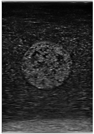
Figure 1 Ultrasound image
Phantoms are devices that can reproduce characteristics in living organisms to validate medical imaging techniques. In fact, ultrasound phantoms have mechanical and acoustic parameters similar to biological tissues, and capable of providing realistic ultrasound images. Their main advantage is their ability to simulate a wide range of geometries, as well as mechanical and acoustic parameters, in the validation of imaging techniques (registration, segmentation, etc.) under realistic and controlled conditions. In most cases, phantoms are designed with mechanical and acoustic properties that come as close as possible to the organs targeted by the developed applications (kidneys, liver, breasts, etc.).
In elastographyDef.“Elastography is a medical imaging modality that maps the elastic properties and stiffness of soft tissue. The main idea is that whether the tissue is hard or soft will give diagnostic information about the presence or status of disease. For example, cancerous tumours will often be harder than the surrounding tissue, and diseased livers are stiffer than healthy ones. The most prominent techniques use ultrasound or magnetic resonance imaging (MRI) to make both the stiffness map and an anatomical image for comparison.”(Wikipedia, https://en.wikipedia.org/wiki/Elastography), for example—a technique used to estimate the mechanical properties of biological tissues for diagnostic purposes—phantoms make it possible to compare results of a tested algorithm with the known properties of the phantom.
Several commercial phantoms already make the reproduction of a wide variety of configurations possible with a high degree of accuracy. However, these phantoms are very expensive. In addition, when it comes to reproducing configurations that may vary throughout the test phases—different geometries, different mechanical and acoustic properties—“home-made” phantoms make more sense.
Several scientific works reported in the literature propose recipes to make phantoms, while characterizing their mechanical and acoustic properties. However, no study provides specific instructions for achieving well-defined mechanical properties. In other words, no study provides a validated predictive model to determine the elastic modulus of a phantom-simulating soft tissue using easily adjustable parameters.
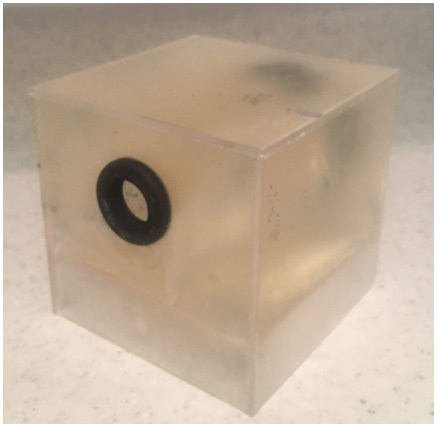
Figure 2: Ultrasound phantom created in a laboratory
Predictive Model of the Elastic Modulus
The study provides a model that predicts the elastic modulus of a phantom, by adjusting easily controllable parameters—gelatin concentration and refrigeration time—while using a simple, fast and inexpensive recipe commonly used in the medical ultrasound community. This research will help systemize phantom design for researchers in the fields of ultrasound and elastography. Depending on the desired elasticity of the phantom, the user can choose the appropriate proportions and manufacturing process using a predictive function. Several normal and pathological soft biological tissues can be represented with the proposed recipe, such as normal breast fat (elastic modulus of 19 kPa), fibrous breast tissue (107 kPa), or normal prostate tissue (69 kPa).
Among other things, the study showed that the elastic modulus of a phantom can be adjusted within a range of 14 kPa to 103 kPa by varying the gelatin concentration in the mixture from 5 g/100 ml to 20 g/100 ml. In addition, the study shows that refrigeration time only increases the elastic modulus from 2 hours to 24 hours, and from 2 hours to 42 hours, after which the elastic modulus decreases. Statistical analysis confirms that gelatin concentration and refrigeration time constitute statistically significant control parameters in the model.
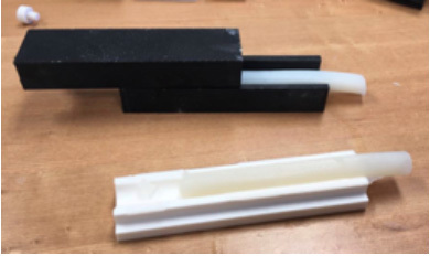
Figure 3: Phantom samples for mechanical characterization
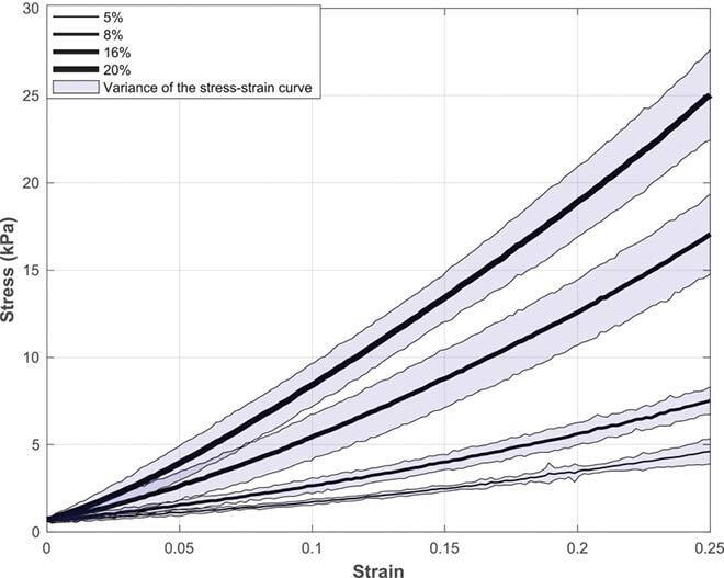
Figure 4: Tensile tests for different gelatin concentrations
The prediction model gives estimates on optimal refrigeration time and gelatin concentration to obtain a target elasticity modulus with a high confidence interval (> 95%). The elastic modulus predictive model was validated on small cylindrical samples (<20 ml) and large-scale phantoms, such as those used in ultrasound and elastography (600 ml). Results showed that the model provides a good prediction of the elastic modulus (error <16%) in both cases.
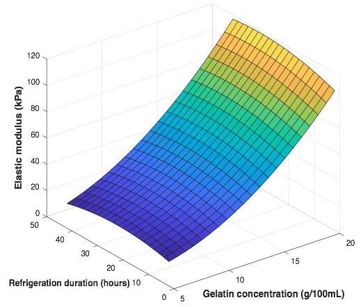
Figure 5: Elastic modulus depending on gelatin concentration and refrigeration time
Regarding the acoustic properties, results of the study showed that increasing the gelatin concentration increases the acoustic attenuation and impedance, while decreasing sound speed in the phantom. Increasing the cooling period increases the attenuation coefficient for all gelatin concentrations.
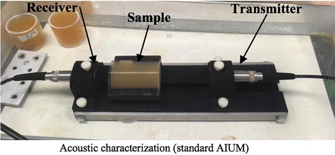
Figure 6: Assembly used for acoustic characterization
The acoustic impedance achieved by the phantom under study is comparable to biological soft tissues. Regarding the estimated attenuation coefficient, results show that only specific configurations of gelatin concentration and refrigeration time can reach values comparable to the gelatin and biological soft tissue phantoms documented in the literature. Finally, with regard to sound velocity, estimated values are lower than those reported for biological soft tissues (1560 m/s on average). However, several additives are proposed in the literature to adjust the acoustic parameters, including sound velocity.
As for the ultrasound image texture obtained by the proposed phantom, it is reproduced using hydrophilic mucilloid fibre (Metamucil, a low-cost product available in any drug store). A statistical study also showed that the Metamucil concentration in the mixture affects the ultrasound image texture.
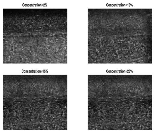
Figure 7: Ultrasound texture obtained at different gelatin concentrations
Conclusion
This study provides a predictive model of elasticity as a function of two easily adjustable parameters: gelatin concentration and refrigeration time. The model can be used as a guide to design phantoms mimicking human tissues and to reach a target modulus of elasticity with a good confidence interval, an important practical contribution to the field. The phantom is produced with a simple, fast and inexpensive recipe using easily available ingredients and can mimic a variety of healthy and diseased biological tissues with a range of elastic properties of 15 kPa to 100 kPa. The acoustic parameters—sound velocity, acoustic impedance—of the phantom are also comparable to biological tissues, providing good quality ultrasound images.
Additional Information
For more information on this research project, please read the following paper:
Dahmani, J.; Laporte, C. Pereira, D.; Bélanger, P. Petit, Y. 2020. “Predictive Model for Designing Soft-Tissue Mimicking Ultrasound Phantoms with Adjustable Elasticity.” IEEE, Volume 67, Issue 4, pp. 715-726.