Automatic Extraction of Vertebral Landmarks from Ultrasound Images

Purchased on Istock.com. Copyright.
The clinical standard for monitoring scoliosis is based on X-ray images. Unfortunately, repeated exposure to X-rays is harmful to the patient’s health. Ultrasound is a radiation-free medical imaging technique that uses ultrasound (US) waves to acquire images. However, interpreting vertebral ultrasound images is difficult because image quality can vary greatly. In response to this challenge, we present a method that automatically locates vertebrae on ultrasound images. Since we validated this as a reproducible approach, it would be possible in time to replace some X-ray examinations with ultrasound scans. Keywords: Ultrasound imaging, spine, vertebrae, scoliosis, anatomical landmarks, automatic detection.
Reducing Radiation in Scoliosis Monitoring
Scoliosis is a 3D deformation of the spine that has the potential of worsening over time. The clinical standard for monitoring its evolution is to perform biplane X-ray imaging of the spine (Figure 1) to estimate the position and orientation of the vertebrae in 3D. Unfortunately, repeated exposure to X-rays is harmful to the patient’s health [1].
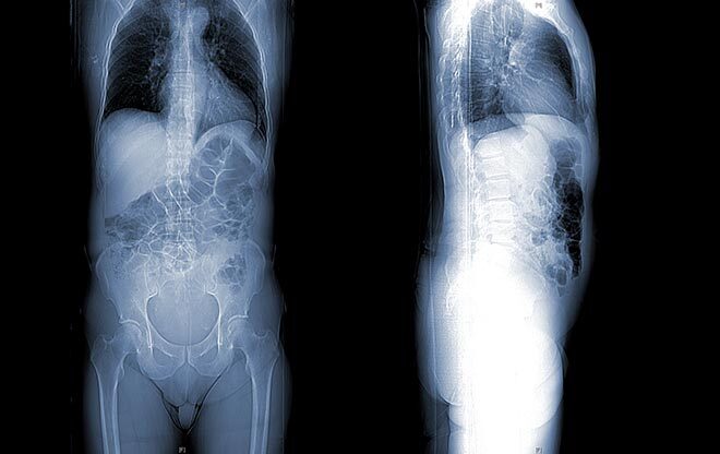
Figure 1: Biplane X-ray images (front + side)
A radiation-free alternative to X-rays is the freehand ultrasound approach [2]. It is a medical imaging technique that combines a standard 2D US probe with a position sensor to acquire 3D images [3]. However, interpreting vertebral ultrasound images is difficult because image contrast can vary greatly during their acquisition. This is why these images are usually interpreted manually by clinicians. The relevance of their interpretation is therefore dependent on their expertise and is time consuming.
Vertebral Landmarks—A New Approach
Our solution to this problem is to automatically extract anatomical landmarks on the vertebrae. On ultrasound scans, a vertebra looks like a W divided into two V’s by a vertical symmetry axis (Figure 2). The connection point between the two V’s corresponds to the spinous process (SP); the top part of the symmetry axis of each V corresponds respectively to the left (LL) and right (RL) laminae of the vertebra. Note that the W-shape is not fully visible and that the contrast does not allow easy and precise localization of its position.
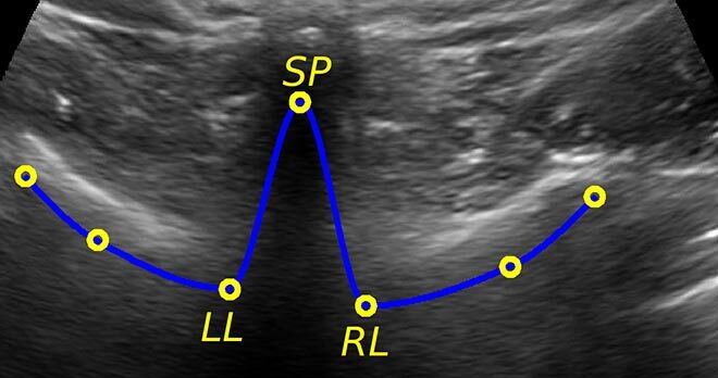
Figure 2: W-shape of the vertebra in blue, with three anatomical landmarks (SP: spinous process, LL: left lamina, and RL: right lamina) in yellow.
Our method is based on this information to ensure the reproducible extraction of one landmark for each anatomical area: SP, LL and RL (Figure 3). To do so, the respective sums of pixel intensities in the rows (right) and columns (top) of the image are computed (in blue) then standardized (in green). Anatomical landmarks are then identified based on the zero crossings (in red).
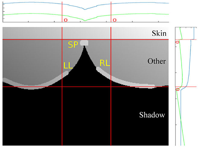
Figure 3: Schematic view of a vertebral ultrasound image with the sums of pixel intensities along rows (right) and columns (top), before (blue) and after (green) standardization.
Validation of the Proposed Method
Our method has been validated through two experiments. First, 3D vertebral ultrasound images of a porcine cadaver were acquired using a freehand approach with a reference frame defined by markers attached to one vertebra. The corresponding 3D volume was also reconstructed using a CT scan as a reference. However, the volume coordinates were expressed with respect to a reference frame that is specific to the CT scan. A geometric transformation was then applied to the coordinates in order to express them within the same reference frame as the ultrasound scans, which produces a CT section corresponding to each ultrasound scan.
Second, images of the vertebrae of several healthy subjects were acquired using 2D ultrasound scanning. Three observers with ultrasound experience manually annotated the vertebral landmarks on each ultrasound image. The average position of the three annotations was taken as a reference.
The positions of the automatically extracted vertebral landmarks—obtained using our method—are very similar to those obtained using the reference methods—CT scan or average manual annotation—i.e. they are less than 2 mm apart, when the ultrasound image is of good quality. Knowing that the manual annotation of an ultrasound image takes about 30 s, while our method takes less than a second to perform, we can conclude that our method represents a reproducible and rapid solution for vertebral ultrasound image analysis.
Conclusion
Our results are promising and suggest that it would be possible in time to replace some X-ray examinations with an ultrasound examination. This would limit the patient’s exposure to radiation and possibly allow for more frequent examinations, ensuring better monitoring of scoliosis and a more favourable treatment for patients.
Additional information
For more information on the research, please read the following paper:
Brignol, A., Gueziri, H. E., Cheriet, F., Collins, D. L., & Laporte, C. (2020). “Automatic extraction of vertebral landmarks from ultrasound images: A pilot study.” Computers in Biology and Medicine, 103838.