June 18, 2026
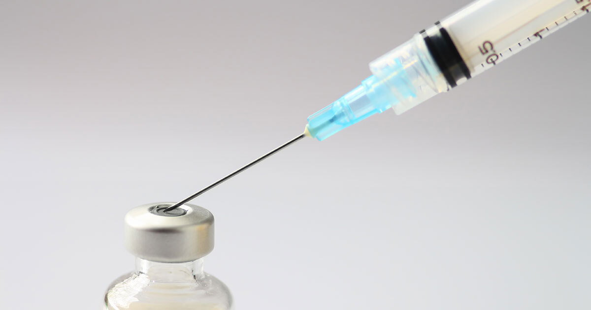
What if serious diseases could be treated without resorting to major surgery, simply by injecting a “smart” material that can act exactly where the body needs it? This is the idea behind the research of Sophie Lerouge, a professor at ÉTS and holder of the Canada Research Chair in Injectable Biomaterials for Cell and Endovascular Therapy. By developing hydrogels and microparticles—whether or not they contain living cells—her team designs innovative solutions for biomedical applications ranging from endovascular embolization to cell therapy and tissue engineering.
Based at the Centre hospitalier de l’Université de Montréal (CHUM) research center, her laboratory stands out as an interdisciplinary approach focused on clinical transfer. It brings together polymer chemistry, materials mechanics, cell biology, and medical practice, all united by a single goal: addressing actual clinical needs through biomaterials engineering.
New injectable embolizing agents
Key to Sophie Lerouge’s research is the embolization of blood vessels. This technique involves intentionally blocking vessels to stop unwanted blood flow, for example, in cases of arteriovenous malformations or internal bleeding, or to cut off the blood supply to certain tumours. Since current embolizing agents fail to meet clinical needs, Sophie Lerouge and her team are developing injectable materials, in liquid form that gel inside the human body when exposed to heat, or in the form of microspheres. Administered via catheter during endovascular procedures, these embolizing agents make open surgery unnecessary, reducing risks and recovery times, and can be combined with bioactive agents. This research is conducted in close collaboration with interventional radiologists at the CHUM, Dr. Gilles Soulez and Éric Thérasse, and has led to several patents, one of which is currently being brought to market by a leading company in the field of endovascular treatments.
Some embolizing particles are designed to release local anesthetics. This is crucial because embolization causes ischemia—a reduction or stoppage of blood flow to a tissue. This lack of oxygen and nutrients can be very painful, especially during procedures such as uterine fibroid treatments.
More recently, the research team has also been exploring drug-loaded magnetic microparticles. Guided remotely by a magnetic field—namely, using MRI—these particles could be directed toward very specific arteries without needing to position the catheter physically at the site of the procedure. This approach paves the way for even more selective and less invasive embolization.
Hydrogels and cell therapy
Professor Lerouge’s second major area of research focuses on cell therapy and tissue engineering. Cell therapy involves injecting living cells—stem cells or immune cells—to repair tissues or fight diseases such as cancer. However, when injected alone, these cells disperse rapidly, have poor survival rates, and often lose their effectiveness.
To address this issue, Sophie Lerouge and her team are developing injectable hydrogel-based matrices. In liquid form at room temperature, these materials can be mixed with cells or drugs and then injected using very fine needles or catheters. Once inside the body, they solidify at body temperature and form a porous structure: a true “home” for the cells.
This porosity is essential. It allows cells to receive sufficient oxygen and nutrients, multiply, interact with one another, and, when necessary, gradually leave the matrix to wield their therapeutic action.
Cancer immunotherapy: localized, long-lasting action
These hydrogels hold particular promise in cancer immunotherapy. The method harnesses the patient’s T cells (sometimes genetically modified) to recognize and attack tumour cells. Existing treatments often rely on systemic injections of billions of cells, with only a tiny fraction actually reaching the tumour, at the cost of significant side effects.
The strategy developed by Sophie Lerouge and her team is different and focuses action exactly where needed. Encapsulating T cells in an injectable hydrogel directed toward the tumour enables the use of far fewer cells while prolonging and enhancing their effectiveness. The cells multiply within the matrix, forming three-dimensional structures, and gradually migrate toward the tumour, attacking it continuously rather than randomly.
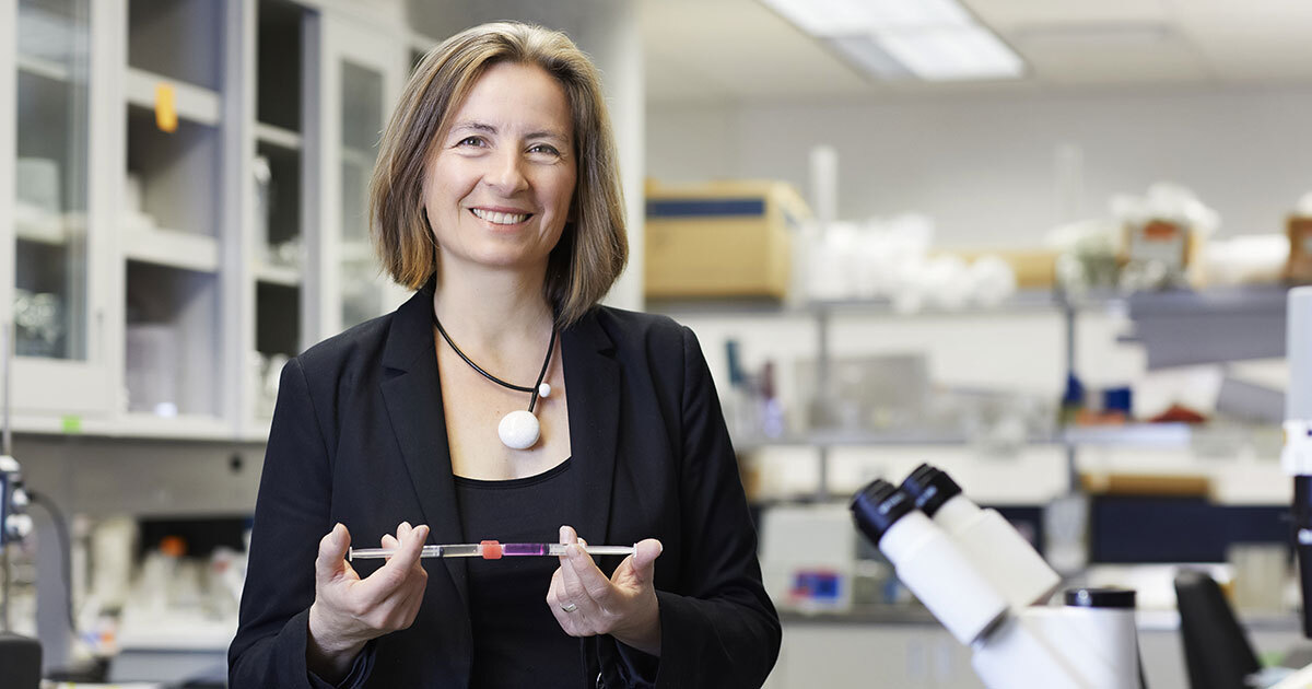
ÉTS professor Sophie Lerouge
Very encouraging results have already been obtained in animal models, showing significant improvement in anticancer activity. In this instance, the material does more than simply transport cells: it regulates their release, survival, and interaction with the tumour microenvironment.
Tissue regeneration using stem cells
The hydrogels developed by the team are also used for stem cell transplantation, namely in ischemic tissues—that is, tissues deprived of sufficient blood supply. Stem cells possess proangiogenic properties: they promote the formation of new blood vessels and the regeneration of damaged tissues.
When injected into a suitable matrix, these cells can survive longer, multiply, and gradually release molecules with beneficial effects. Here again, the main challenge is striking a balance: retaining enough cells for a lasting effect while allowing the diffusion of their therapeutic factors.
Toward 3D bioprinting
Beyond direct therapeutic applications, the hydrogels developed by Sophie Lerouge and her team also play a role in 3D bioprinting. This technology, which involves printing structures containing living cells, could transform biomedical research and, in the longer term, regenerative medicine.
The challenges are many: hydrogels tend to collapse, cell survival is difficult to maintain during printing, and biocompatibility must be balanced with structural precision. To address these issues, the researcher is developing, in collaboration with her colleague Ali Ahmadi, new bio-inks and innovative bioprinting methods to improve both structural resolution and cell viability.
In the short term, these advances will enable the creation of micro-organs or tissue models that are more realistic than Petri dish cultures, allowing for more reliable and personalized testing of drug efficacy and toxicity. In the longer term, they could contribute to the production of tissues and even implantable organs.
Collaborative and educational research
Located within a hospital setting, Sophie Lerouge’s laboratory also provides an exceptional training environment. Students are exposed to a wide variety of technologies, from materials design to preclinical feasibility testing, in a highly multidisciplinary context.
“Our projects always stem from a real clinical need,” explains the researcher. “Our role is to use engineering and biomaterials to overcome actual medical challenges.” A philosophy that sets collaboration, innovation, and societal benefits at the heart of research.
Sophie Lerouge is a member of itechsanté, the ÉTS research institute for innovation in health technologies. To learn more about the institute, its mission, themes, flagship projects, and more, visit itechsanté