
Areas of research
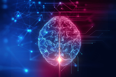
The biomedical field combines multiple technologies that produce large volumes of information pertaining to the functioning of the human organism. The integrated management of this information is carried out in a variety of ways, including multimodal fusion, which involves taking many types of information into consideration in order to arrive at the optimal solution to a problem.
The team of Catherine Laporte, Jean-Marc Lina and Rita Noumeir propose new data analysis methods, as much for helping doctors in their decision-making, and thus improving the care provided to patients, as for promoting the emergence of scientific discoveries related to human health through inter-sector collaborations.
Examples of the type of data being analyzed:
- EEG (electroencephalography) and MEG (magnetoencephalography) information from electromagnetic brain imaging among epileptic patients;
- Pairing of brain oscillations observed using EEG and localized using MEG (sleep study);
- NIRS (diffuse optical imaging) and MEG information in cognitive brain imaging;
- Fusion of MRI-X-ray-ultrasound for monitoring treating scoliosis (non-invasive acquisition of rachis volumes in the upright position);
- Videos acquired in the visible and infrared spectrum for decision support in pediatric intensive care;
- Non-structured notes available in the electronic health record for identification and automatic extraction of critical clinical situations;
- Ultrasound scanning of the vocal tract, video recordings of the face and acoustic signals associated with the production of speech for the articulation study;
- Eye tracking and inertial data acquired in conjunction with ultrasound images for the purpose of studying hand-eye coordination among sonographers.
The areas of research of each of our researchers are detailed below:
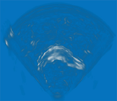
Non-invasive scoliosis monitoring techniques using ultrasound scanning
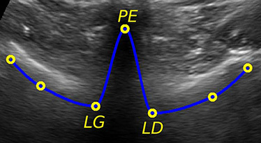
Adolescent idiopathic scoliosis is a complex 3D deformity of the rachis that affects between 1% and 3% of the population. Among those affected, 1 in 200 suffers from a deformity that requires treatment due to its progression. There is currently no reliable method for predicting which scoliotic deformities present a risk of progression, so all suspected cases are monitored via a series of X-rays acquired semi-annually throughout the rapid growth period. Unfortunately, the cumulative exposure to X-rays significantly increases the incidence of certain types of cancer. LATIS is working in collaboration with the Sainte-Justine University Hospital Research Centre (Dr. Marie-Claude Miron, Dr. Hubert Labelle, Dr. Stefan Parent and Dr. Farida Cheriet) to develop non-invasive techniques for monitoring the shape of the spine based on freehand 3D ultrasound scanning, an acquisition protocol that allows for large-volume reconstruction from a series of 2D ultrasound images of a localized area thanks to a position sensor on the scanner, or even specialized image matching algorithms. In addition, techniques based on a robust and global variation of ultrasound elastography are being studied for improving the characterization of the role that back muscles play in the progression of the condition. Our current projects focus on machine learning methods for guiding an ultrasound scanner in the acquisition of rachis volumes along an optimized trajectory based on the quality of the volume obtained, which will have a significant impact on the adoption of this technology in the practical world.
Analysing the mechanism of tongue movement using ultrasound scanning
The characterization of the shape and movement of the tongue is important to the study of the production of healthy and pathological speech. Because it is mobile, non-intrusive and inexpensive, ultrasound scanning has proven to be very useful in this context. LATIS is working in collaboration with Professor Lucie Ménard (Linguistics Department, UQAM) and Professor Steven Lulich (Audiology and Speech Sciences Department, Indiana University Bloomington) to develop the tools required to analyze the articulatory dynamics of the tongue, and specifically for segmenting the tongue in 2D and 3D and tracking its movements in an ultrasound video, and for characterizing how these shapes and movements differ from one population to another (e.g.: among healthy adults compared to young children). Our current projects focus on the application of these concepts to the longitudinal study of articulatory movements with respect to speech, but also with respect to the development of the speaker.
Human-machine interaction in medical imaging
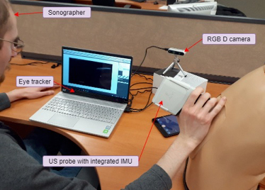
More than any other imaging method, ultrasound scanning relies on the knowledge and experience of the practitioner acquiring the information, which is reflected mainly in the quality of the image produced, but also in the abundance of contextual information in their interpretation in a clinical setting. This highlights the relevance of the human-machine interaction in the treatment and analysis of the images produced through ultrasound scanning. Working in collaboration with Professor Michael John McGuffin (Software and IT Engineering Department, ÉTS) LATIS has developed methods for segmenting images that allow for the contour of a targeted anatomical structure to be extracted in an interactive and fluid manner, which is a particularly onerous problem due to the many artefacts that decrease the quality of ultrasound images. We are currently expanding the application context of these human-machine interactions to include the acquisition of the images themselves, or in other words, the operator’s manipulation of the probe. This is more important than ever before, because there are now more affordable miniaturized ultrasound scanners available on the market, making this technology accessible to new users. A measurement system based on eye-tracking and the analysis of trajectories and images is being developed for the purpose of ultimately studying the visuo-motor coordination of expert sonographers. The knowledge that is acquired will then be used to design navigation tools to facilitate learning among new users.
Signal processing in electroretinography
Retinal imaging is essentially an automatic imaging of the back of the eye to measure the architecture of the vascular network. Working in collaboration with the Ophthalmology Department at the McGill University Health Centre (Dr. Pierre Lachapelle) for the past five years, LATIS has been developing methods for functional characterization of the back of the eye using electroretinography measurements (measuring the electric potential at the surface of the cornea). These methods, which have been evaluated in animal models and longitudinal clinical databases, have shown incredible clinical potential. LATIS continues to develop tools for analyzing the electric potential measured at the cornea, focusing on two specific aspects: the localization of functional anomalies on the retina and analysis of the ERG signal at rest (multifractal analysis).
Electromagnetic imaging of brain activity
There are three methods for non-invasive measurement of bioelectrical brain activity:
- Electroencephalography (electric potential at the scalp) or EEG, the most common method;
- Magnetoencephalography (magnetic field at the exterior of the head) or MEG, with six facilities in Canada, including two in Montréal;
- Diffuse optical imaging (diffusion and absorption of infrared light) or NIRS, the most recent.
We are developing entropic methods for localizing brain activity using these external measurements. These digital methods are now among the most reliable techniques for determining regions of neural activation (applications in epilepsy and sleep studies among an aging population). LATIS is one of the most active research groups in terms of developing innovative methods related to electromagnetic brain imaging in humans. The current focus is on localization of neural connectivity networks and the dynamic of interactions between these functional networks.
Analyzing rhythms and the absence of rhythms
The majority of clinical physiological activity biomarkers rely on the presence of spontaneous and transient oscillations. LATIS has developed an expertise in analyzing rhythms, especially within the context of measurable electrical brain activity, including electroencephalography. These rhythms are studied for their synchronization among different sites, and for their localized interactions. They are primarily analyzed within the context of sleep among normal populations (aging study) and clinical populations (epilepsy, Parkinson’s, REM sleep disorders, etc.). In addition to the rhythmic aspects of physiological signals, LATIS is conducting research into the arrhythmic component of signals, such as multifractal analysis using wavelets. From a different perspective, rhythms (especially respiration) are also studied in terms of measurable movements during sleep, thanks to a matrix of pressure sensors.
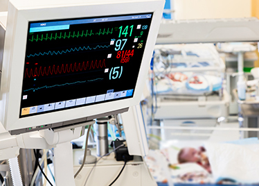
Research Chair on the Development and Validation of Clinical Decision Support Systems using Artificial Intelligence
Professor Noumeir holds the prestigious Research Chair on the Development and Validation of Clinical Decision Support Systems using Artificial Intelligence. She is working closely with Dr. Philippe Jouvet and intensive care physicians at Sainte-Justine University Hospital (CHU Sainte-Justine) to develop computerized clinical decision support systems.
The goal is to analyze wide-ranging medical data using methods based on artificial intelligence in order to develop a computerized clinical decision support system (CDSS). A CDSS is a set of algorithms and interfaces that analyzes and summarizes information to guide health professionals or managers in their decision-making. The system is fed various medical data that are processed through artificial intelligence algorithms. The data include the following:
- Clinical data: Demographic data, clinical assessments, diagnoses, physical exam notes, lab results, medical images, specialized test results;
- Therapeutic data: Doses and administration of medication, ventilator data, data from organ failure machines;
- Organizational data: Number and severity of diseases, patient distribution, distribution and workload of nurses;
- Image and video data: Acquisition of RGB, depth and infrared images.
These data are massive, varied and fully cover a patient’s progress over time. The objective is to use these data to help caregivers with their decision-making.
Electronic patient health record
The electronic health record has been the recipient of major investments. It will help to improve the quality of care and increase the efficacy and efficiency of the health care system. However, the technological challenges are enormous. Some of the problems that are of interest to LATIS are the compression of medical images with a view to facilitating archiving and transfer time over telecommunication networks, the extraction of structure from non-structured medical data, with the goal of automatic comprehension of medical document, and modelling of the flow of patients and medical information to improve care and reduce waiting lists.
Intelligent video surveillance
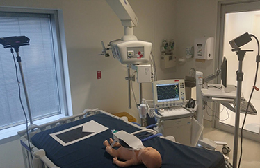
LATIS is interested in intelligent video surveillance. A number of collaborative research projects have received funding through the NSERC, FRQS, MITACS, Prompt, TransMedTech, IVADO and the private sector. Specific collaborations include working with ASI on the detection and recognition of activity and working with CHU Sainte-Justine in connection with diagnostic support for intensive care patients. We use a number of RGB-D (Kinect) and infrared cameras to acquire and combine videos. We use methods based on artificial intelligence to process these videos to detect specific situations of interest (e.g.: suicide attempt or overdose) in real time, or to measure specific parameters that cannot be measured using other methods (e.g.: level of consciousness, respiratory rate or volume).